Visual Analytics
Group leader: Lei Zheng
Projects:
-
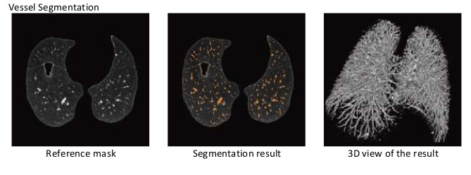
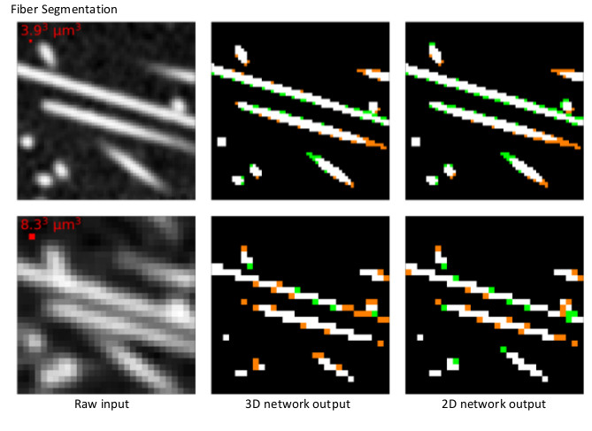
Image Segementation with Deep Learning
To identify thin tubular structures at low resolution and to distinguish them from each other from 3D CT scans are of high interest in medical image processing for vessel segmentation [1] and industrial image processing for fiber segmentation [2][3]. Most of the top-scoring methods are based on cost functions derived from the eigenvalues of the Hessian on the image. Instead of using handcrafted filters, our idea is to automatically compute a set of convolution filters particularly tuned to the dataset using deep learning architectures. The framework of fully convolutional architecture with residual units is applied to such tasks. A residual unit consists of two convolutional layers followed by a batch normalization and a non-linearity layer. Different model types like slice-wise 2D model and 3D model with different number of convolutional layers were explored.
Researchers: Tomasz Konopczyński, Lei Zheng
-
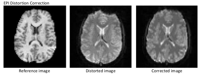
Multi-Modal Image Registration
Multi-modal image registration is a key element for image analysis allowing to benefit from the combined information of different image modalities. Such ill-posed problem can be formulated to minimize a target function with an intensity-based similarity metric and a regularization term. As the intensities of multi-modal images are usually in different scales, mutual information (MI) and its variation normalized mutual information (NMI) are the choices of intensity-based similarity metric. However, they suffer from a relatively flat landscape of function values. Different strategies should be explored to ensure the robustness of this high dimensional ill-posed inverse problem. For distortion correction of echo-planar imaging (EPI) data, a novel anisotropic regularization functional was introduced as the regularization term [4]. Furthermore, to integrate additional geometry information in the cost function, a metric homotopy was introduced to generalize the multi-metric registration approach [5].
Researchers: Daniel Glodeck, Lei Zheng
-
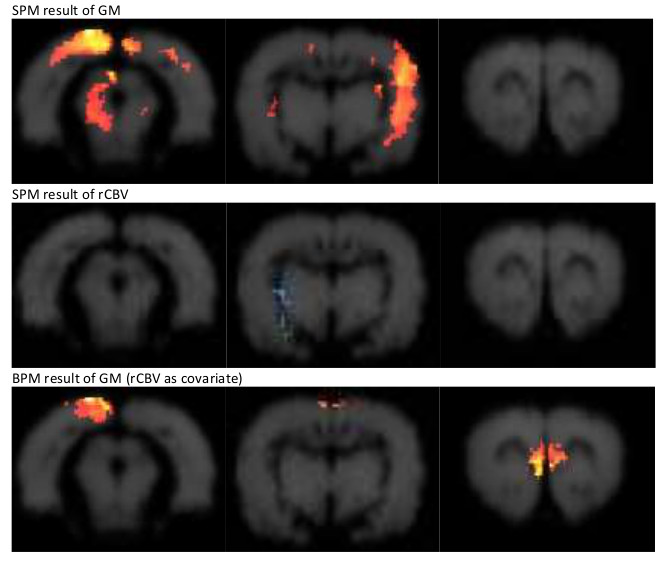
Voxel-Based Morphometry
Voxel-based morphometry (VBM) is a non-invasive method used to investigate brain structural changes. In this method, high-resolution 3D MRI images are analyzed to measure gray matter (GM), white matter (WM) and cerebrospinal fluid (CSF) volumes. The obtained GM maps can be used to visualize changes in the brain tissues. To understand the VBM findings, it is essential to explore the sensitivity of VBM to the influencing effects. With the development of multimodal analysis tools, VBM was investigated together with regional cerebral blood volume (rCBV) changes as condition-wise imaging covariates in ANCOVA analysis, and no evidence of significant interaction between both was found [6].
Researcher: Lei Zheng
Other projects:
-
Facial Landmark Detection
-
Vessel Segmentation and Applications
-
Brain Segmentation and Applications
References
. 2016. “Automated Multiscale 3D Feature Learning For Vessels Segmentation In Thorax Ct Images”. Ieee Nuclear Science Symposium And Medical Imaging Conference. Ieee Nuclear Science Symposium And Medical Imaging Conference. doi:10.1109/NSSMIC.2016.8069570.
. 2017. “Reference Setup For Quantitative Comparison Of Segmentation Techniques For Short Glass Fiber Ct Data”. In 7Th Conference On Industrial Computed Tomography (Ict 2017). 7Th Conference On Industrial Computed Tomography (Ict 2017). Leuven, Belgium. http://www.ndt.net/events/iCT2017/app/content/Paper/26_Konopczy-ski_Rev4.pdf.
. 2018. “Fully Convolutional Deep Network Architectures For Automatic Short Glass Fiber Semantic Segmentation From Ct Scans”. In 8Th Conference On Industrial Computed Tomography (Ict 2018). 8Th Conference On Industrial Computed Tomography (Ict 2018). Wels, Austria. http://www.ndt.net/article/ctc2018/papers/ICT2018_paper_id120.pdf.
. 2016. “Distortion Correction Of Epi Data Using Multimodal Nonrigid Registration With An Anisotropic Regularization”. Magnetic Resonance Imaging 34. Magnetic Resonance Imaging: 127 - 136. doi:http://dx.doi.org/10.1016/j.mri.2015.10.032. http://www.sciencedirect.com/science/article/pii/S0730725X15002696.
. 2018. “Potential Of Metric Homotopy Between Intensity And Geometry Information For Multi-Modal 3D Registration”. Z Med Phys. 28 (4). Z Med Phys.: 334. doi:10.1016/j.zemedi.2018.01.004. https://www.sciencedirect.com/science/article/pii/S0939388917301435.
. 2016. “Influence Of Regional Cerebral Blood Volume On Voxel-Based Morphometry”. Nmr Biomed 29 (6). Nmr Biomed: 787-95. doi:10.1002/nbm.3519.